Hip Muscles Diagram : Hip Anatomy Eorthopod Com. Smartdraw includes 1000s of professional healthcare and anatomy chart templates that you can modify and make your own. The hip joint is a ball and socket synovial type joint between the head of the femur and. The strong muscles of the hip region also help to hold the hip joint together and prevent dislocation. Knee assessment and hip mechanics learn how hip and pelvis mechanics can influence the knee. This article will introduce the muscles in each group and touch on their origin, insertion, function, and innervation.
ads/bitcoin1.txt
Ligaments are soft tissue structures that connect bones to bones.a joint capsule is a watertight sac that surrounds a joint.in the hip, the joint capsule is formed by a group of three strong ligaments that connect the femoral head to the acetabulum. The four muscle of the quadriceps all extend the lower leg, and the rectus femoris additionally can flex the thigh at the hip. The hip joint is one of the most flexible joints in the entire human body. This article will introduce the muscles in each group and touch on their origin, insertion, function, and innervation. The four groups are the anterior group, the posterior group, adductor group.
What Muscles Does Running Work Running Works Leg Muscles And The Core from i.insider.com The posterior and lateral muscles the posterior and lateral muscles are also referred to as the muscles of the buttocks. This is the largest of the three compartments of the thigh. The hip flexors are several muscles that bring your legs and trunk together in a flexion movement. The quadriceps group of four muscles. Ligaments, tendons, and muscles play an important role in the function of the hip. Large ligaments, tendons, and muscles around the hip joint hold the bones (ball and socket) in place and keep it from dislocating. The hip joint is one of the most flexible joints in the entire human body. The muscles in the hip are responsible for the movement of the hip and, by proxy, the leg.
The four muscle of the quadriceps all extend the lower leg, and the rectus femoris additionally can flex the thigh at the hip.
ads/bitcoin2.txt
Extension, flexion, adduction, and abduction. Hip flexion is maximal with a high, forward kick that brings the leg above the level of the waist. Ligaments are soft tissue structures that connect bones to bones.a joint capsule is a watertight sac that surrounds a joint.in the hip, the joint capsule is formed by a group of three strong ligaments that connect the femoral head to the acetabulum. What that means in practical terms is that they turn the leg outward. The femur may also rotate around its axis about 90 degrees at the hip. The hip joint is a ball and socket synovial type joint between the head of the femur and. Muscles and tendons also serve to protect joints. The strong muscles of the hip region also help to hold the hip joint together and prevent dislocation. The posterior and lateral muscles the posterior and lateral muscles are also referred to as the muscles of the buttocks. Related posts of muscles of the lower back and hip diagram muscle recovery anatomy. The hip muscles work together to carry out 4 different types of movement: These muscles run from the lower spine and pelvis, join together, then attach by a tendon to the upper thigh. These muscles can be grouped based upon their location and function.
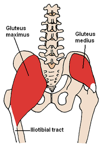
The muscles which make up this group include the gluteus minimus, the gluteus maximus, the gluteus medius, and the tensor fasciae latae. Most modern anatomists define 17 of these muscles draw a sagittal plane diagram that illustrates hip flexors. Iliopsoas muscle, a hip flexor muscle that attaches to the upper thigh bone. The diagram shows the posterior (rear) view of the buttock. This is the largest of the three compartments of the thigh.
Hipflexor Muscle Anatomy Hip Anatomy Hip Muscles Anatomy from i.pinimg.com The many muscles of the hip provide movement, strength, and stability to the hip joint and the bones of the hip and thigh. Hip flexion is maximal with a high, forward kick that brings the leg above the level of the waist. This is the largest of the three compartments of the thigh. These muscles run from the lower spine and pelvis, join together, then attach by a tendon to the upper thigh. The pelvic floor muscles provide foundational support for the intestines and bladder. Smartdraw includes 1000s of professional healthcare and anatomy chart templates that you can modify and make your own. When, in pilates or another fitness class, the instructor says turn your leg out in the hip socket. As you can see from the diagram to the right, there are many muscles and tendons that make up the hip and buttocks region.
The gluteus maximus can be seen at the top, cut away to expose the plantar view of foot:
ads/bitcoin2.txt
Related posts of muscles of the lower back and hip diagram muscle recovery anatomy. Hip pain can sometimes be caused by diseases and conditions in other areas of your body, such as your lower back. The piriformis is the horizontal muscle in the center of the picture running over the top of the sciatic nerve. The hamstring muscles, which originates mostly from the ischial tuberosity and insert on the tibia/fibula, also assist with hip extension. These muscles run from the lower spine and pelvis, join together, then attach by a tendon to the upper thigh. More commonly, our hips flex to a 90° angle when we sit. Muscles of thigh and the hip diagram. The diagram shows the posterior (rear) view of the buttock. The hip joint is a ball and socket synovial type joint between the head of the femur and. Rectus femoris and the sartorius can cause some movement in the hip joint but these muscles primarily move the knee, and not generally classified as muscles of the hip. Muscles and tendons also serve to protect joints. The strong muscles of the hip region also help to hold the hip joint together and prevent dislocation. The hip muscle diagram below shows a number of the muscles we will be discussing in the next sections.
Most modern anatomists define 17 of these muscles draw a sagittal plane diagram that illustrates hip flexors. The hip muscles encompass many muscles of the hip and thigh whose main function is to act on the thigh at the hip joint and stabilize the pelvis.without them, walking would be impossible. They allow you to move your leg or knee up towards your torso, as well as to bend your torso forward at the hip. These are often divided into four groups according to their orientation around the hip joint: Hip flexion is maximal with a high, forward kick that brings the leg above the level of the waist.
Muscles Of The Hip Wikipedia from upload.wikimedia.org You can strain or tear your hip flexor muscles through sudden movements or falls. These are often divided into four groups according to their orientation around the hip joint: They allow you to move your leg or knee up towards your torso, as well as to bend your torso forward at the hip. There are various hip flexor muscles that all work to. Tensor fascia lata trigger point in it band and hip pain dr perry details the tensor fascia late trigger point that cause hip pain and it band syndrome hip injuries hip disorders take a look at some mon and not so mon hip injuries and disorders find tests and details on different problems of the hip and clinical trials The strong muscles of the hip region also help to hold the hip joint together and prevent dislocation. The diagram shows the posterior (rear) view of the buttock. The iliopsoas muscle is a powerful hip flexor that runs across the top of the hip joint and works to pull the knee up and off the ground.
The four groups are the anterior group, the posterior group, adductor group.
ads/bitcoin2.txt
Muscles and tendons also serve to protect joints. The hip flexors can be found connecting the top of the femur, which is the largest bone in the body, to the lower back, hips, and groin. The piriformis is the horizontal muscle in the center of the picture running over the top of the sciatic nerve. As you can see from the diagram to the right, there are many muscles and tendons that make up the hip and buttocks region. When, in pilates or another fitness class, the instructor says turn your leg out in the hip socket. The view on the left has the rectus femoris cut away to show the vastus intermedius which is below it. Hip flexion is maximal with a high, forward kick that brings the leg above the level of the waist. The muscles work together to enable movement and keep the hip in alignment. Rectus femoris muscle, one of the quadriceps muscles on the front of your thigh. When you flex your hip, you move the leg forward. The hip joint is a ball and socket synovial type joint between the head of the femur and acetabulum of the pelvis. The hip joint is a ball and socket synovial type joint between the head of the femur and. They allow you to move your leg or knee up towards your torso, as well as to bend your torso forward at the hip.
ads/bitcoin3.txt
ads/bitcoin4.txt
ads/bitcoin5.txt
0 Response to "Hip Muscles Diagram : Hip Anatomy Eorthopod Com"


0 Response to "Hip Muscles Diagram : Hip Anatomy Eorthopod Com"
Post a Comment